QuASAR program shrinks equipment and removes temperature constraints for high-resolution sensing and imaging at nano-scale
May 2, 2013
In science, many of the most interesting events occur at a scale far smaller than the unaided human eye can see. Medical researchers might realize a range of breakthroughs if they could look deep inside living biological cells, but existing methods for imaging either lack the desired sensitivity and resolution or require conditions that lead to cell death, such as cryogenic temperatures. Recently, however, a team of Harvard University-led researchers working on DARPA’s Quantum-Assisted Sensing and Readout (QuASAR) program demonstrated imaging of magnetic structures inside of living cells. Using equipment operated at room temperature and pressure, the team was able to display detail down to 400 nanometers, which is roughly the size of two measles viruses. For a sense of scale, see: http://learn.genetics.utah.edu/content/begin/cells/scale/.
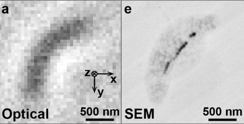
The Harvard QuASAR team’s technique is described in a Nature paper titled “Optical magnetic imaging of living cells.” Essentially, the researchers used imperfections in diamond known as nitrogen-vacancy (NV) color centers to function as high-precision probes of the magnetic fields produced by living magnetotactic bacteria—organisms that contain magnetic nanoparticles. Using an array of these NV color centers engineered at specific points and density within a diamond chip, the researchers were able to locate the magnetic structures in each bacterium and construct images of the magnetic fields they produced.
The team’s findings have several potential applications and could lead to additional areas of study:
- In principle, this technique would enable detailed, real-time observation of internal cellular processes, such as cell death, evolution and division, and how cells are affected by disease.
- The researchers’ measurements are directly applicable to studying formation of magnetic nanoparticles in other organisms, which is of interest for contrast enhancement in magnetic resonance imaging (MRI), and has been linked to neurodegenerative disorders.
- Formation of magnetic nanoparticles has been proposed as a mechanism for magnetic navigation in higher organisms.
In a related development, two separate teams of QuASAR researchers, led by the University of Stuttgart in Germany and IBM’s Almaden Research Center, developed a nano-scale magnetometer that enables magnetic resonance imaging (MRI) with sufficient resolution to measure as few as 10,000 protons in a volume of only 125 cubic nanometers, which approaches the level of individual protein molecules. Previous MRI technologies, even when stretched to their limits, have not allowed for resolution beyond a few micrometers because of distorting factors like background magnetic noise. The QuASAR teams’ new technique, dubbed nano-MRI, overcomes this limitation by using a single NV color center embedded close to the surface of a diamond chip to measure nuclear magnetic resonance signals. It can be used to measure the magnetic field at a single point on a structure, or scan across the surface to image the structure by measuring multiple points. The work is described in two papers in the February 1, 2013 edition of Science: “Nanoscale Nuclear Magnetic Resonance with a Nitrogen-Vacancy Spin Sensor” and “Nuclear Magnetic Resonance Spectroscopy on a (5-Nanometer)3 Sample Volume.”
The nano-MRI technology offers the additional benefit of working at room temperatures, eliminating the need for expensive cryogenic equipment. Conversely, traditional MRI uses bulky machinery that frequently requires cryogenic cooling.
The University of Stuttgart and IBM’s work on QuASAR could potentially offer a range of future medical benefits and capabilities:
- Support future drug development by facilitating increased understanding of the structure of proteins.
- Enable detailed, three-dimensional mapping of biological molecules, with sufficient sensitivity to identify specific elements. This information could streamline assessment of inhibitor drugs against naturally occurring and bioengineered viruses.
- Enable measurement of the magnetic field of firing neurons.
“In QuASAR we are building sensors that capitalize on the extreme precision and control of atomic physics. We hope these novel measurement tools can provide new capabilities to the broader scientific and operational communities,” said Jamil Abo-Shaeer, DARPA program manager. “The work these teams are doing to apply quantum-assisted measurement to biological imaging could benefit DoD’s efforts to develop specialized drugs and therapies, and potentially support DARPA’s work to better understand how the human brain functions.”
All three efforts were conducted as basic research. Future work may include attempts to: increase the sensitivity of the measurement devices by moving them even closer to the organisms to be measured; embed nano-diamonds with NV centers into living cells for in-vitro studies to measure magnetic fields and temperature; and enable NV-assisted magnetic-field imaging of tagged bio-molecules.
# # #
Associated images posted on www.darpa.mil may be reused according to the terms of the DARPA User Agreement, available here: http://www.darpa.mil/policy/usage-policy.
Tweet @darpa
